
It’s a term used to describe a series of rare conditions that affect the development of a baby’s skull.
During the development of an infant, folds of membranes called Interdigitations form between the bones to create a structure commonly known as a Suture.
The term Suture refers to a hinge of bone edges or joints united by a thin layer of soft tissue.
There are different types of Sutures in the human skulls, and these joints usually close at various stages of an individual’s life depending on the type and its location.
Types of Sutures
- Metopic Suture, it refers to the only cranial structure that naturally closes up during infancy. It runs mid-line of the frontal bone and usually fuses between the third and ninth month after birth.
- Sagittal Suture, it is the serrated joint located between the two parietal bones of the skull. More often than not, this suture fully closes between the ages of 30 and 40 years old.
- Coronal Sutures, it refers to the fibrous connective tissue joint located between the frontal bone and the parietal bones. It fuses between the 18 and 24 months after birth.
- Lambdoid Sutures, also known as the lambdoidal sutures, are the junction between the parietal bones and the occipital bone. They start fusing between 25 to 30 years and fully close at the age of 70 years old.
- Squamosal Suture, also known as the squamous suture, is the cranial joint located between the temporal and parietal bones bilaterally. It begins to fuse by the age of 24 and fully closes between 30 and 40 years of age.
- Frontal Sphenoid Suture, also known as the sphenofrontal suture, is the vertical cranial suture between the sphenoid and temporal bones bilaterally. It usually closes by the third month after birth.
Definition
Craniosynostosis refers to the early fusion of one or more sutures in an infant’s skull before birth or after delivery.
These joints or gaps (the sutures) need to remain open for the brain of any child to grow and develop normally and healthily, and gradually close or fuse as the human development goes on.
However, in the case of the infants affected by this condition, those joints in the skull fuse prematurely, which prevents the perpendicular expansion of the child’s head.
That forces the head to compensate by growing parallel to the closed sutures, which results in the malformation of the skull.
The abnormal shape of the head often differs from case to case depending on the affected suture.
Types of Craniosynostosis
Each potential case of Craniosynostosis gets classified into several kinds based on which particular sutures are fusing prematurely.
Following such reasoning, people often acknowledge four types of Craniosynostosis, which get listed here below:
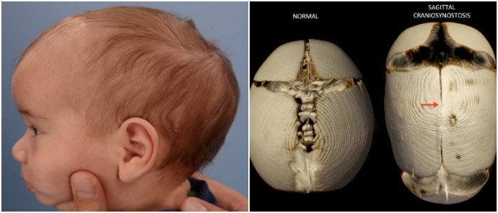
Sagittal Craniosynostosis
It’s also known as Sagittal Synostosis. Several doctors regard this condition as the most common type of Craniosynostosis.
Sagittal Craniosynostosis consists of the premature closure of the sagittal suture, which results in the skull growing longer but narrower than usual.
Coronal Craniosynostosis
This condition occurs due to the fusion of one of the two coronal sutures that run from the top of the ear to the top of the skull.
It results in the infant developing a flat forehead with the abnormal growth focusing on the affected side.
This malformation also affects some facial features, which is why patients may display a raised eye socket and a crooked nose.
In some cases, both coronal sutures get fused, and the child ends up developing a flat and prominent forehead and brow.
Metopic Craniosynostosis
It’s also known as Trigonocephaly. This condition is a rare type of Craniosynostosis that features the premature fusion of the metopic suture.
That results in the infant developing a forehead with a triangular appearance and having the back part of the head widened.
Lambdoid Craniosynostosis
It’s also known as Lambdoid Synostosis. Some doctors regard this condition as the rarest type of Craniosynostosis.
In this particular case, the fusion occurs at the lambdoid suture, which results in affected children developing a flattened head at the back.
However, not every infant with a flattened head at the back has Lambdoid Craniosynostosis. Sometimes, a child may present such feature due to lying on their backs for prolonged periods.
If such is the case, the child has a condition that medical experts call “Positional Plagiocephaly,” also known as Flat Head Syndrome.
A General Practitioner can help a family to determine if their baby has a case of Craniosynostosis or Flat Head Syndrome.
Causes
Researchers are still trying to pinpoint the reason behind the development of Craniosynostosis.
After taking a closer look to 20% of the cases featuring a single suture Craniosynostosis, some theorize that a mutation that changes the genes may be the cause of these conditions.
On the other hand, some evidence suggests the premature fusion of the sutures occurs when the bones of the skull get restricted in the womb, not allowing for movement from the growing brain.
In some cases, single suture Craniosynostosis occurs as the result of a Craniofacial Syndrome, a disorder caused by a genetic mutation.
Symptoms
Something that occurs in all types of Craniosynostosis is what medical experts call an increased Intracranial Pressure (ICP).
ICP refers to a measurement of the pressure inside the skull and thus in the brain tissue and cerebrospinal fluid that cushions and surrounds the brain and the spinal cord.
When fusion occurs to only one suture, there’s a 15% probability that a child may experience increased ICP.
That may result in persistent headaches, vision issues, and some developmental problems in the patient’s academic abilities.
In most cases, patients that display such symptoms don’t suffer from increased ICP. However, if left untreated, the condition may lead to other symptoms such as the ones listed here below:
- Vomiting
- Sluggishness or unresponsiveness
- The difficulty at following a moving object
- Problems to hear clearly
- Struggling to breathe properly
Other signs are pretty apparent at birth, such as the misshaped head as well as a strange feeling or disappearing fontanel on the infant’s skull.
A raised and hard ridge tends to form along the affected suture. And in many cases, the growth of the head doesn’t match the development of the rest of the body.
Sometimes, a baby may appear asymptomatic at birth, but after the first month of life, the condition may become more noticeable.
Diagnosis
The malformation of the head it’s a typical symptom that even the family of the patient can notice.
However, the people who are more qualified to determine or identify cases of Craniosynostosis are pediatric neurosurgeons or plastic surgeons.
Some pediatricians may even rely on the evaluation provided by a craniofacial specialist. Still, the diagnosis of this condition often includes the tests listed here below:
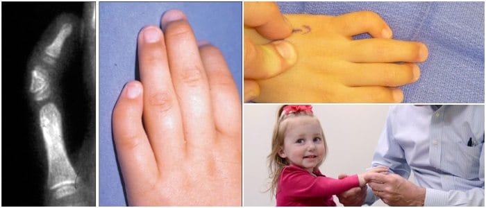
- Physical Exam, which involves the medical expert feeling the baby’s head for abnormalities such as suture ridges, and look for facial deformities.
- Computed Tomography (CT) scan, it uses rotating X-ray machines to create cross-sectional images of the body. It can show whether any sutures have fused in the baby’s skull.
- Genetic Testing, it is a medical test used to determine changes in chromosomes, genes, or proteins. It can help doctors to identify any underlying genetic disorder.
Treatment
In mild cases of Craniosynostosis, some doctors may recommend the use of a specially moulded helmet to correct the shape of the infant’s skull and assist the growth of the baby’s brain.
However, in most cases, medical experts consider that surgery is the best possible chance for the patient to grow and develop healthily.
Such a matter is also relevant for the child’s mental health as an unusual head shape can negatively impact a kid’s personality, self-esteem, and social interactions.
The goal of this medical procedure is to expand the volume of the skull, which would relieve the ICP on the brain. Some people know this surgical operation as “Cranial Vault Remodeling.”
This treatment consists of remodeling or reshaping the bones of the patient’s head to expand and enlarge the space within the skull that contains the brain.
Such a procedure is meant to give the brain more room to grow while attempting to restore a more natural appearing shape of the skull.
The specialists that perform the surgery are the craniofacial surgeon and the pediatric neurosurgeon working as a team to reduce the amount of time a child requires under anesthesia.
Medical experts recommend performing this surgical treatment before the child reaches a year of age. However, the best time for the procedure may depend on the affected sutures and their condition.